Part 1. Nerve History.
Sensory peripheral nerves send information to the central nervous system from internal organs or from external stimuli.
https://secure.health.utas.edu.au/intranet/cds/histoprac/Practicals/6.Nerve.html
1. Sketch and label three structures on each slide, including these:
On Slide 16, identify the Node of Ranvier and myelin sheath.
On Slide 23, identify the nerve and nerve fascicles.
2. Compare the bundling of nervous tissue with that of muscle tissue. Include a discussion of connective tissue arrangement.
To the left, is a bundle of the nervous tissue. It is very similar to the structure of the muscle tissue. It comprises of numerous long cells "bundled" closely next to each other.
And to the right, is an image of the muscle bundle. The parallel fibers that tacked together.
The human body is composed of just four basic kinds of tissue: nervous, muscular, epithelial, and connective tissue. Connective tissue is the most abundant, widely distributed, and varied type. Muscle contraction begins when the nerve fiber releases the neurotransmitter acetylcholine into the synaptic cleft. Acetylcholine moves across the synaptic cleft and binds to receptors on the muscle fiber. This indirectly initiates an action potential, a change of electrical charge at the membrane that is similar to events in a neuron.
Sketch the relationship between a sensory neuron, an interneuron, and a motor neuron (as in Figure 11.2). Label each part and state the function of each type of neuron.
The brain processes stimuli from the environment through its five senses: sight, taste, touch, sound, and smell. Think of an example of how your brain processed each of these types of stimuli, as you got ready for school this morning. Is there a measurable difference between the rate at which one can process sensory information obtained visually, orally, and tactually?
I know that all three of these stimuli are equally important to our survivor. For me, I believe, the sight and sound are the best processors. We all have the different reaction times to each of these stimulants and it all depends on our individual nerve receptors and transmitters. Or simply, sensitivity.
Materials:
2. Compare the bundling of nervous tissue with that of muscle tissue. Include a discussion of connective tissue arrangement.
To the left, is a bundle of the nervous tissue. It is very similar to the structure of the muscle tissue. It comprises of numerous long cells "bundled" closely next to each other.
And to the right, is an image of the muscle bundle. The parallel fibers that tacked together.
The human body is composed of just four basic kinds of tissue: nervous, muscular, epithelial, and connective tissue. Connective tissue is the most abundant, widely distributed, and varied type. Muscle contraction begins when the nerve fiber releases the neurotransmitter acetylcholine into the synaptic cleft. Acetylcholine moves across the synaptic cleft and binds to receptors on the muscle fiber. This indirectly initiates an action potential, a change of electrical charge at the membrane that is similar to events in a neuron.
3. Why is the distal part of a peripheral nerve narrower than its proximal part?
Proximal parts of the peripheral nerves are wide because they sectioned out with the Schwan's Cells. They are protected by myelin sheaths. The distal areas of the nerves narrower, because they do not have such sheaths, and they are the first receptacles of the signal.
4. The myelin sheath of a nerve accepts biological dye easily because of the abundance of lipids in the membranes wrapping around the axon. In cross section, the myelin sheaths look like donuts. The adipose cells may stand out as they contain preserved fat droplets that stain dark brown-black.
See this link http://image.shutterstock.com/display_pic_with_logo/1350394/159810476/stock-photo-myelin-sheath-of-peripheral-nerve-fibers-stained-with-osmium-tetroxyde-light-microscope-micrograph-159810476.jpg
A deficiency in the lipids of the myelin sheath can diagnose diseases. What is one such disease? What are the symptoms?
Lack of lipids can cause the disformity of the myelin sheath. This leads to A disease called Multiple Sclerosis or MS. There is a variety of symptoms, but the most profound are muscle weakness, visual impairment, and urinary incontinence. Also, similar but much more rare ALS (Amyotrophic Lateral Sclerosis) known as Lou Gehrig's disease. Progressive weakness of skeletal tissue causes the person to die, because of inability to support the inner organs, of suffocation.
Motor peripheral nerves carry information from the central nervous system to organs, muscles, and glands.
5. View the motor end plate side (100x), sketch and label three structures. Motor end plate image: https://secure.health.utas.edu.au/intranet/cds/histoprac/images/H1685x100.jpg
Part 2 Anatomy of a Neuron
Sketch the relationship between a sensory neuron, an interneuron, and a motor neuron (as in Figure 11.2). Label each part and state the function of each type of neuron.
Part 3 Reaction Time Rulers
The brain processes stimuli from the environment through its five senses: sight, taste, touch, sound, and smell. Think of an example of how your brain processed each of these types of stimuli, as you got ready for school this morning. Is there a measurable difference between the rate at which one can process sensory information obtained visually, orally, and tactually?
I know that all three of these stimuli are equally important to our survivor. For me, I believe, the sight and sound are the best processors. We all have the different reaction times to each of these stimulants and it all depends on our individual nerve receptors and transmitters. Or simply, sensitivity.
Materials:
Ruler or meter stick
Partner
Record keeping:
Visual Stimulus
|
Auditory Stimulus
|
Tactile Stimulus
|
||||
Person 1
|
Person 2
|
Person 1
|
Person 2
|
Person 1
|
Person 2
|
|
Trial 1 L |
18
|
19
|
5
|
18
|
15.5
|
13
|
Trial 1 R |
11
|
17
|
13.5
|
3
|
13.5
|
13.5
|
Trial 2 L |
17
|
18
|
7
|
16.5
|
14
|
11.5
|
Trial 2 R |
11
|
15
|
13
|
8
|
11.5
|
13
|
1. Which type of stimulus did you respond to more quickly? Your partner? Why do you think that is?
I have responded more quickly with my right hand to the AUDITORY stimulus. My partner has responded to the same stimulus but with his left hand. I think this is the result of a better sensitivity to the sound.
2. Which type of stimulus did you respond to more slowly? Your partner? Why do you think that is?
The VISUAL stimulus proved to be the slowest one for me, and for my partner. My vision is not perfect, but I think there may be a "broken" link between by visual receptors and neurological response.
3. Explain why a message moving along nerve pathways takes time.
The time we are talking about is the milliseconds, but it does take time for the "message" to reach the receiver. Electrical signal activates sensory neuron and creates an action potential, the exchange of Na(+) and K(+) defuse the stored Ca(2+) into axon bulb. This "wakes up" the neurotransmitters and synaptic transmission occurs. The "message" has been delivered to the local region and we have a reaction to the distress. (like pulling a hand away from the fire).
4. Explain the process of a reflex.
An action that is performed as a response to a stimulus and without conscious thought. And: (of an action) performed without conscious thought as an automatic response to a stimulus.
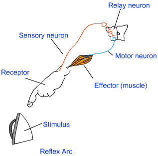
The path through which nerves signals; involved in a reflex action; travel is called the reflex arc. The following flow chart shows the flow of the signal in a reflex arc.
Reflex action is a special case of involuntary movement in voluntary organs. When a voluntary organ is in the vicinity of a sudden danger, it is immediately pulled away from the danger to save itself. For example; when your hand touches a scalding electric iron, you move away your hand in a jerk. All of this happens in a flash and your hand is saved from the imminent injury. That is an example of reflex action.

5. Was your hypothesis supported?
The sight has proved to be a good stimulus for me, but the sound, for my surprise, have failed me.
Part 4 Divisions of the Peripheral Nervous System
Complete this table:
Somatic division Autonomic division
Somatic division Autonomic division
Sympathetic
|
Parasympathetic
|
||
Function
|
Serves skeletal muscles
|
Fight or flight; arouses body to deal with situations involving physical activity, mental alertness.
|
Relaxes the body, Promotes digestion and other basic functions.
|
Neurotransmitter
|
Acetilcoline.
|
Norepinephrine.
|
Acetylcholine.
|
Works Sited:
"Types of Neurons Home - Hook AP Psychology 3B." Types of Neurons Home - Hook AP Psychology 3B. Web. 21 Oct. 2015.
"Muscle Fibres, SEM." - Stock Image P154/0196. Web. 23 Oct. 2015.
"Muscle." - Biology Encyclopedia. Web. 24 Oct. 2015.
"Control and Coordination." Class Ten Science Control and Coordination Human Brain. Web. 25 Oct. 2015.
"Google." Google. Web. 25 Oct. 2015.


I can't believe this. A great testimony that i must share to all Multiple Sclerosis is patient in the world i never believed that their could be any complete cure fo rMultiple Sclerosis i saw people's testimony on sites of how DR JACOB helped them with the cure of Multiple Sclerosis i had to try it too and you can't believe that in just few weeks i started using it all my pains stopped so I had to go and meet my doctor to see if still have Multiple Sclerosis but to my greatest surprise I was negative, thank you you so much www drjacobherbal . online/ WhatsAPP +1 (336) 962-8303
ReplyDelete