Monday, October 26, 2015

Week 10. The Digestive and Endocrine Systems Lab


My virtual daily menu

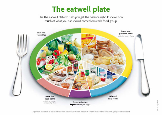
Have you noticed that instead of Food Pyramid we now have an Eat Well Plate? Here it is!


Journal Entries:


1) Explain why the menu of food choices you made is nutritious, meaning the food choices follow the government's recommendations for Calories and nutrients.

I believe that the choices I have made are nutritious because I have included all of the recommended food groups. I also have no Trans Fats, and I think it is a good thing.  I also have plenty of protein intake, so I will have enough energy to go about my daily routine 

2)  How does a 2 000 Calorie daily menu compare to your recommended Calorie intake amount on the chart? What would you do to your 2 000 Calorie menu to make it better match your recommended Calorie intake?

From the chart, I was recommended the 2,000 calory diet. So in that region I am doing pretty good. Because it is an average of what I do eat, some days I have more or less depending on the amount of the physical activity. Looks like I can afford some additional calories so a chocolate chunk will be a great treat as a snack. 

3) How can you use the Nutrition Facts label to make healthy decisions?

The obvious reason not to eat something if it has more than 50% of any of one nutrient. If there is too much fat or salt, it is going to hurt you sooner or later. But, if there is not enough of something, like o% fat or 0% calcium. It is also not good because you are not getting the needed "supply". So depends on what you eat, you can use these labels as your guide and make the decision based on your personal needs, and not because some commercial told you to eat it. 

4) Did the menu you created contain the foods you like to eat? Explain why or why not.

Absolutely. I would not be choosing something I will choke on (as can't eat it because it is too gross). Everybody should eat what they like. Otherwise, it will be no "fun" to eat. The moderation is the key. I love caramel ice cream, but I will not be eating it all day long. Because I also like the bread and the meat, and the salads. I can have different foods every day, and I will not get bored with it. Also, the snacks can bring great variety to your daily meals. Different fruit, different kind of nuts, different veggies. 

5) Does any of the items you selected provide more than 30% in any nutritional area? If so, which foods and which nutrients? Why did you choose those foods?

I have all of the areas above the 30% mark. Except the trans fats one. That one has 0%.
As I went back and retried my diet, I don't have any one ingredient that is over 30% on anything. But combining them all brings it up. So if I chose cheese and the refried beans, I don't think I am overdoing it on anything because tomorrow I will not do not have the same food. Plus, I chose these foods because I love to eat them. 

6) What did you notice about the nutrients in fast foods?

Most of the offered fast foods contain almost half of the daily value of one ingredient or many. Most have too much of salt, and almost all have lots of fats. 

7) What did you notice about the food group icons in your Daily Meals area? Did you favor a particular food group? Explain.

All of the icons correspond mostly to the five food groups. Grains, Dairy, Protein, Vegetables, and Fruits. The subcategory Oils, Fats and Candies seem to bundle the others all together, so it doesn't matter if you chose a tomato or a refried beans, you got yourself a vegetable! 
I think I favored the Vegetable group. All other groups are equally present. I love variety; that is why I have a little of everything on my plate.


Works Sited:

"The Digestive and Endocrine Systems." The Digestive and Endocrine Systems. Web. 26 Oct. 2015.

"Food Pyramid & Portion Sizes." SoFaB Kids Camp. Web. 26 Oct. 2015.


Wednesday, October 21, 2015

Week 9. Nervous System Lab.


Part 1. Nerve History.


Sensory peripheral nerves send information to the central nervous system from internal organs or from external stimuli.

https://secure.health.utas.edu.au/intranet/cds/histoprac/Practicals/6.Nerve.html

1. Sketch and label three structures on each slide, including these:

 On Slide 16, identify the Node of Ranvier and myelin sheath.


On Slide 23, identify the nerve and nerve fascicles.


2. Compare the bundling of nervous tissue with that of muscle tissue. Include a discussion of connective tissue arrangement.


To the left, is a bundle of the nervous tissue. It is very similar to the structure of the muscle tissue. It comprises of numerous long cells "bundled" closely next to each other.


And to the right, is an image of the muscle bundle. The parallel fibers that tacked together. 
The human body is composed of just four basic kinds of tissue: nervous, muscular, epithelial, and connective tissue. Connective tissue is the most abundant, widely distributed, and varied type. Muscle contraction begins when the nerve fiber releases the neurotransmitter acetylcholine into the synaptic cleft. Acetylcholine moves across the synaptic cleft and binds to receptors on the muscle fiber. This indirectly initiates an action potential, a change of electrical charge at the membrane that is similar to events in a neuron. 

3. Why is the distal part of a peripheral nerve narrower than its proximal part?


Proximal parts of the peripheral nerves are wide because they sectioned out with the Schwan's Cells. They are protected by myelin sheaths. The distal areas of the nerves narrower, because they do not have such sheaths, and they are the first receptacles of the signal.

4. The myelin sheath of a nerve accepts biological dye easily because of the abundance of lipids in the membranes wrapping around the axon. In cross section, the myelin sheaths look like donuts. The adipose cells may stand out as they contain preserved fat droplets that stain dark brown-black. 

See this link http://image.shutterstock.com/display_pic_with_logo/1350394/159810476/stock-photo-myelin-sheath-of-peripheral-nerve-fibers-stained-with-osmium-tetroxyde-light-microscope-micrograph-159810476.jpg

A deficiency in the lipids of the myelin sheath can diagnose diseases. What is one such disease? What are the symptoms?

Lack of lipids can cause the disformity of the myelin sheath. This leads to A disease called Multiple Sclerosis or MS. There is a variety of symptoms, but the most profound are muscle weakness, visual impairment, and urinary incontinence. Also, similar but much more rare ALS (Amyotrophic Lateral Sclerosis) known as Lou Gehrig's disease. Progressive weakness of skeletal tissue causes the person to die, because of inability to support the inner organs, of suffocation.

Motor peripheral nerves carry information from the central nervous system to organs, muscles, and glands.

5. View the motor end plate side (100x), sketch and label three structures. Motor end plate image: https://secure.health.utas.edu.au/intranet/cds/histoprac/images/H1685x100.jpg

Part 2 Anatomy of a Neuron 

Sketch the relationship between a sensory neuron, an interneuron, and a motor neuron (as in Figure 11.2). Label each part and state the function of each type of neuron.


Part 3 Reaction Time Rulers 

The brain processes stimuli from the environment through its five senses: sight, taste, touch, sound, and smell. Think of an example of how your brain processed each of these types of stimuli, as you got ready for school this morning. Is there a measurable difference between the rate at which one can process sensory information obtained visually, orally, and tactually?

I know that all three of these stimuli are equally important to our survivor. For me, I believe, the sight and sound are the best processors. We all have the different reaction times to each of these stimulants and it all depends on our individual nerve receptors and transmitters. Or simply, sensitivity. 

Materials:

Ruler or meter stick
Partner

Record keeping:
   Visual Stimulus
   Auditory Stimulus
   Tactile Stimulus
Person 1
Person 2
Person 1
Person 2
Person 1
Person 2

Trial 1 L
    18
     19
     5
     18
     15.5
     13

Trial 1 R
    11
     17
    13.5
      3
     13.5
     13.5

Trial 2 L
    17
     18
     7
     16.5
     14
     11.5

Trial 2 R
    11
     15
     13
      8
     11.5
     13


1. Which type of stimulus did you respond to more quickly? Your partner? Why do you think that is?

I have responded more quickly with my right hand to the AUDITORY stimulus. My partner has responded to the same stimulus but with his left hand. I think this is the result of a better sensitivity to the sound. 

2. Which type of stimulus did you respond to more slowly? Your partner? Why do you think that is?

The VISUAL stimulus proved to be the slowest one for me, and for my partner. My vision is not perfect, but I think there may be a "broken" link between by visual receptors and neurological response.

3. Explain why a message moving along nerve pathways takes time.

The time we are talking about is the milliseconds, but it does take time for the "message" to reach the receiver. Electrical signal activates sensory neuron and creates an action potential, the exchange of Na(+) and K(+)  defuse the stored Ca(2+) into axon bulb. This "wakes up" the neurotransmitters and synaptic transmission occurs. The "message" has been delivered to the local region and we have a reaction to the distress. (like pulling a hand away from the fire).

4. Explain the process of a reflex.

An action that is performed as a response to a stimulus and without conscious thought. And: (of an action) performed without conscious thought as an automatic response to a stimulus.
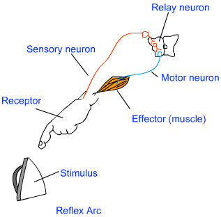
The path through which nerves signals; involved in a reflex action; travel is called the reflex arc. The following flow chart shows the flow of the signal in a reflex arc.

Reflex action is a special case of involuntary movement in voluntary organs. When a voluntary organ is in the vicinity of a sudden danger, it is immediately pulled away from the danger to save itself. For example; when your hand touches a scalding electric iron, you move away your hand in a jerk. All of this happens in a flash and your hand is saved from the imminent injury. That is an example of reflex action.


5. Was your hypothesis supported?

The sight has proved to be a good stimulus for me, but the sound, for my surprise, have failed me. 

Part 4 Divisions of the Peripheral Nervous System

Complete this table:


                          Somatic division                                                  Autonomic division


Sympathetic
Parasympathetic
Function
  Serves skeletal muscles
 Fight or flight; arouses body to deal with situations involving physical activity, mental alertness.
 Relaxes the body, Promotes digestion and other basic functions.
Neurotransmitter
Acetilcoline.
 Norepinephrine.
 Acetylcholine.


Works Sited:


"Types of Neurons Home - Hook AP Psychology 3B." Types of Neurons Home - Hook AP Psychology 3B. Web. 21 Oct. 2015.

"Sciencephotolibrary." Science Is Everything (Nerve Bundle. Coloured Scanning Electron...). Web. 23 Oct. 2015.

"Muscle Fibres, SEM." - Stock Image P154/0196. Web. 23 Oct. 2015.

"19 Breathtaking Out-Of-This-World Photos." Diply. Web. 23 Oct. 2015.

"Do Motor Neurons Have the Same Structure as Sensory Neurons?Frequently Asked in." Do Motor Neurons Have the Same Structure as Sensory Neurons? Web. 23 Oct. 2015.

"Muscle." - Biology Encyclopedia. Web. 24 Oct. 2015.

"Control and Coordination." Class Ten Science Control and Coordination Human Brain. Web. 25 Oct. 2015.

"Google." Google. Web. 25 Oct. 2015.

Johnson, Michael D. Human Biology: Concepts and Current Issues. 7th ed. San Francisco: Pearson Benjamin Cummings, 2014. Print.

Wednesday, October 14, 2015

Week 8. Lab 7. Immune System


Part 1. Erythrocytes and Leukocytes. 

(I can sketch and label the cells by hand, but I think for the sake of better viewing, I will be using copies of the slides from this website: http://www.bu.edu/histology/m/t_periph.htm )


These are "stained" blood cells, done for the better viewing.
The numerous "donut" looking cells are erythrocytes, or as commonly known - the red blood cells.


The labeled slide illustrates the different cells and identifies them.

We know of five types of the leukocytes, the white blood cells. They are separated into two major categories: granular leukocytes (granulocytes) and agranular leukocytes (agranulocytes). The difference between the two is that stained agranulocytes do not show their granules (vesicles) and granulocytes do.


The three types of granulocytes are: 

Basophils- release the histamine to attract blood plasma to the injured area.

Eosinophils- defend the body against parasites, such as worms. The eosinophils bombard the "enemy" with digestive enzymes. They also release special chemicals that moderate the severity of the allergic reactions. 

Neutrophils- attacks  large bacteria and some fungi. They are the first ones to combat infections.


The two types of agranulocytes are:


Lymphocytes- classified into two types, B and T. Their names are based on the area where they mature, B cells in a bone marrow and T cells in the thymus gland. The B cells are parents to the plasma cells that produce antibodies which defend us against microorganisms and other foreign invaders. The T cells target and destroy specific threats such a bacteria, viruses, and cancer cells. 

Monocytes- work against viruses and certain bacterial parasites. They differentiate into the macrophages that "eat up" the invaders and cellular debris.


Let's look at some of them. 


These are monocytes, they are quite larger than a red blood cells and have a pretty big nucleus, with the rest of its area occupied by cytoplasm. (Notice that there are no visible organelles, this is agranulocyte).
As you can see here, the nucleus takes more that half of the monocyte's body.


And these are eosinophils
 ( the one on the right side, the one on the left side is a neutrophil).
Also much bigger than a red blood cells. You can see the two lobes of a nucleus and the organelles - granulocyte.


Part 2. Lymphatic System.


One of the lymphatic organs are the tonsils. 
We can see the formation of lymphatic follicles, the crypt of a tonsil, and the overall shape and size of it.


The lymphatic system is physiologically and anatomically relate to the cardiovascular system. 

The Lymphatic system helps maintain the value of the blood. It transports the fats and fat-soluble vitamins, absorbed by the digestive system, to the cardiovascular system. Lymphatic vessels, which resemble the veins, have the walls which contain the one-way valves, and they acquire same help from the skeletal muscles, allowing the constrictions and pressure changes. Beginning at the lymphatic capillaries, merging to form the lymphatic vessels, which further merge into larger and larger vessels eventually creating two major ducts: right and thoracic. These ducts join the subclavian veins near the shoulders, thereby returning the lymph to the cardiovascular system. As you can see in the drawing bellow, the two systems closely follow the same pattern and exchange their contents continuously. One helps the other with their individual functions and keep each other at a best performing levels.


Part 3. Nonspecific Body Defenses. 

1. Name and explain three ways the integumentary system provides the first line of defense.            

The integumentary system, in other words - our skin, provides the first line of defense. The outer layer, the epidermis, continuously being replaced. As the dead cells fall off, so does the bacteria that desired to "enter". Plus, our sweat glands produce natural antibiotic called dermcidin, that can kill a range of harmful bacteria. 
2. Explain the protective role of cilia. From what primary tissue type do cilia arise?

Cilia, the epithelial tissue that have a hair-like projections that beat constantly in a wave-like  motion to sweep mucus upward into our throat, from where we get rid of it by coughing or swallowing it. 

3. Define and sketch the process of phagocytosis.


(I apologize for my artistic inability. This is why I use pictures and other methods to illustrate the topic)

4. Name and sketch two cell types that perform phagocytosis.

Phagocyte and Lysosomes perform the phagocytosis. (The neutrophils and macrophage cells perform this process also.)

5. Describe the process involved in the inflammatory response. Include all chemicals and cell types.

The injured tissue  release the chemicals that "call" in the mast cells that release histamine (basophils also release the histamine) Histamine promotes dilation of the blood vessels which allows the entry of additional proteins and removal of the damaged and destroyed cells. Complement proteins "mark" the bacteria for phagocyte's easy detection.


6. Explain and sketch the mechanism by which complement kills bacteria.


Complement proteins create the holes on the bacterial cell's walls. Excess diffusion of salt and water swell it up and eventually it bursts. 


Part 4. Specific Body Defenses.

1. What is the major histocompatibility complex?      

A unique set of proteins on the cell's surface of an individual's immune system recognize the "marked" cells and do not attack them. These "self"-markers are known as Major Histocompatibility Complex (MHC) proteins. 
                                                                    
2. Describe and sketch the basic structure of an antibody. How many different types of antibodies do you have in your body?

We have 5 identified types of antibodies. Antibody consists of four peptide chains linked together to form a Y shape. Only the receiving ends of each antibody differentiates them. Each one is designed to receive the specific antigen. 


3. Describe and sketch clonal expansion.

When antigen fragment is found by the inactive T cell, The T cell activates and undergoes mitosis, then it quickly begins to produce clones of identical T cells. 


4. How does interferon operate?

Cells that become infected by viruses secrete a group of proteins called interferons. Interferons then diffuse themselves to the nearby healthy cells, binds their cell membrains together and stimulates the healthy cells to produce proteins that interfere with a synthesis of the viral proteins. This makes harder for the viruses to infect the protected cells. 

5. What is the difference between cell mediated and antibody mediated immune response?

Antibody-mediated immune response: The B cells produce the proteins that binds and neutralizes certain antigens. Then it releases them into the lymph, blood stream, and tissue fluid, where they circulate through the body until the need for their action. They protect against viruses and bacteria that are soluble in blood and lymph. 

Cell-mediated immune response: The T cells are dependent on the actions of the other T cells. T cells directly attack the intruder cells and help coordinate other aspects of the immune response. They protect against parasites, bacteria, viruses, fungi, cancerous cells, and cells perceived as foreign. 

6. Name the cells involved in the cell-mediated immune response.

T cells

7. Name the cells involved in the antibody mediate immune response.

B cells

8. Explain the difference between passive immunization and active immunization and give an example of each.

An active immunization is when the vaccine prevents an infectious disease by activating the body’s production of antibodies that can fight off invading bacteria or viruses.

If there is enough time, the active vaccination is preferable. Keep in mind that passive immunizations provide only short-term protection that often lasts just a few weeks before the antibodies are worn down and removed from the bloodstream. By contrast, active immunizations can produce antibodies that last a lifetime. 

Passive immunization, in which antibodies against a particular infectious agent are given directly to the child or adult, is sometimes appropriate. These antibodies are taken from a donor and then processed so the final preparation contains high antibody concentrations. At that point, they are given in the vein or by a shot to the patient.

Passive immunization is often used in children and adults who have weakened immune systems or may not be good candidates for routine vaccinations for other reasons. It can be used with people who haven’t been vaccinated against a disease to which they’ve been exposed. For example, the passive rabies immunization (rabies immune globulin) is commonly used after a certain type of wild animal bites a child. Passive immunizations for hepatitis A (gamma globulin) may be helpful for people traveling to a part of the world where hepatitis A is common. They are typically given before children or adults leave on their trip. These are used less now that there is a vaccine for hepatitis A.


Works cited:

"Medical Histology -- Peripheral Blood Cells." Medical Histology -- Peripheral Blood Cells. Web. 14           Oct. 2015. <http://www.bu.edu/histology/m/t_periph.htm>.


Web. 16 Oct. 2015. <http://www.bioon.com/book/biology/Biological                                 Diversity/BioBookANIMORGSYS.html>.


Web. 17 Oct. 2015. <https://clinicalscienceblogyaseen.files.wordpress.com/2013/04/fig-3-     inflammatory-response2.jpg>.

Wikipedia. Wikimedia Foundation. Web. 17 Oct. 2015. <https://en.wikipedia.org/wiki/Antibody>.


"PSA Poster: Antibiotic Resistant Gonorrhea." PSA Poster: Antibiotic Resistant Gonorrhea. Web. 17 Oct. 2015. <http://www.slideshare.net/KevinHugins/psa-poster-antibiotic-resistant-g>.


Nature.com. Nature Publishing Group. Web. 18 Oct. 2015.       <http://www.nature.com/icb/journal/v89/n1/fig_tab/icb2010117f1.html>.

"Immunizations: Active vs. Passive." HealthyChildren.org. Web. 18 Oct. 2015. <https://www.healthychildren.org/English/safety-prevention/immunizations/Pages/Immunizations Active-vs-Passive.aspx>.

Web. 18 Oct. 2015. <http://imgarcade.com/1/active-immunity/>.

Tuesday, October 6, 2015

Week 7. Lab 6. Blood

This week we can have some fun and play a game!

Go ahead and open this link, 
I promise you will be happy to help the people in need. 


I played this game a few times, just because it was kind of fun. 

I did get confused with an AB Rh- patient and tried to give him an O+ blood. Oh boy, he did not like that at all! 

All other patients were successfully treated and looked pretty happy. 

I liked how easy it was to identify the type of blood. This is the most important task in the determination of which type of blood can patient receive. Because all types A, B, AB, and O can receive the O blood and all of them compatible with the Rh -, people who have the O Rh- blood type are called the universal donors. The AB Rh+ carriers can receive any other type of blood and thus called the universal receivers. The only "picky" ones are the O-, they can receive only  the O- blood. 

Here is a very easy to read chart that can help in remembering blood compatibility:


The white blood cells consist of: 

a.  Basophils are granulocytes that function to release heparin that inhibits blood clotting. They also release histamine to cause inflammation.

b. Eosinophils are granulocytes that function to kill certain parasites and help control allergic reactions.

c. Lymphocytes are agranulocytes that function to produce antibodies that act against specific foreign substances.

d. Monocytes are agranulocytes that leave the bloodstream to function as macrophages that phagocytize foreign particles.

e. Neutrophils are granulocytes that function to phagocytize foreign particles. 


Work cited:

"The Blood Typing Game." - about Blood Groups, Blood Typing and Blood Transfusions. Web. 6 Oct. 2015. <http://www.nobelprize.org/educational/medicine/bloodtypinggame/>.


 Web. 6 Oct. 2015. <https://highered.mheducation.com/sites/dl/free/0072919329/65712/ch14_EOC.doc>. 


Thursday, October 1, 2015

Week 6. Chicken Wing Dissection Lab.

Lab 5.

Materials

Chicken wing (Gallus domesticus)


· If you do not normally purchase these, ask someone in the meat department of your favorite store to purchase just one.

Buffalo Wild Wings only had the boneless specimens and the Safeway butcher couldn't separate the big package to give me just one wing. So, I had to buy the whole chicken, it was less expensive than purchasing the big pack of wings. (Oooohhhh, the trouble I had to go through :) )


Examination gloves, if you have and want to use
Tray or plate
Dissecting tools (small knife, scissors, tweezers)
Paper towels, soap
Magnifying tool, if you have one

Procedure


1. Put on gloves. Thoroughly rinse and dry chicken wing. Place it on a plate or tray.


2. Examine the outside skin tissue. Record at least 4 observations of the chicken skin. (1 point)

  • The skin appears to be loose.
  • There are visible hair/feather follicles that make the surface of the skin lumpy.
  • In addition to hair/ follicle bumps, there are other uneven clumps under the epidermal layer of the skin. 
  • It is white in color.

3. Using scissors, knife, and tweezers, work slowly and carefully to cut the skin and peel it away from the muscle below. Notice the clear connective tissue that holds the skin to the muscles. The probe may be the most effective tool. As you peel off the skin, you may need to cut away some connective tissue.

4. Record at least 4 observations of connective tissue. (1 point)

  • The connective tissue is loosely attached to the skin.
  • It also connected to the muscle.
  • It appears to be very fragile.
  • It is transparent. 
5. Take a photo (.5 point)


6. What specific type of connective tissue is this? (.5 point)

  • It has all of the classifications of loose connective tissue. 

7. Observe the yellowish clumps of fat tissue found outside the skin. Record at least 2 observations of the fat. (.5 point)

  • The fat feels tough, as of in a solid form. (Suturated)
  • It's located sporadically across the surface of the skin. 

8. What is the biological term for the type of cells that store fat? (.5 point)

  • The biological name for this type of the fat cells is adipocytes.

9. Name at least 2 functions of this fat. (1 point)

  • It is an energy storage.
  • It also provides an insulation. 

10. Observe bundles of muscle tissue surrounding the bones. Separate the bundles of muscles by separating them out with your fingers. Begin by inserting your thumb into the muscle by pushing through the connective tissue covering the muscle. It will give way at the natural separations between the muscle bundles.


11. Viewing through a magnifying tool, if you have one, (but still do if you don’t) describe 2 characteristics of the arrangement of the muscle bundles as you see them here. (.5 points)

  • Each muscle bundle is enclosed in connective tissue. (Fascia) 
  • Each one is connected to the joints by an individual tendon and to the bone by the ligament.  

12. Using your textbook or another reference, sketch a representation of the muscles starting with the muscle cell and ending with the whole muscle. Cite your source! (3 points)


http://www.student.loretto.org/humanbiology/BioLinks/chp4/slide11.htm


13. What type of muscle tissue are you viewing? (.5 point)

  • This is a skeletal muscle.

14. Name the function of this type of muscle tissue. (1 point)

  • Its function is to allow the movement of the bones. 

15. Name 2 two characteristics of this muscle tissue. (.5 point)

  • This muscle can contract, upon receiving the signal.
  • It also can relax.

16. Nerves are thin, threadlike, white strands found between the muscle and the nearest bone. Remove a single muscle by cutting the tendons and peeling the muscle away from the bone. Look for the nerve in your specimen. The texture is much different from a tendon or bone. It is rather slippery. Did you find one? (.5 points)
  • I did!
17. Take a photo of the muscle and nerve or only the muscle, if you did not find the nerve. (.5 point)


18. The strong, shiny, white cords of tendons hold the muscle to the bones. Some of these tendons pulled away from the bone as you separated the muscle bundles.

Observe with a magnifying tool, if you have one, describe, and sketch

a. the attachment of a tendon to muscle. (.5 point)
  • Thin, tough, white-ish Tendon becomes more and more like the muscle, it branches out with numerous strands, changes the color,  and completely merges with the muscle itself. 
b. the attachment of a tendon to the bone. (.5 point)
  • Tendon extends its thin fibers and attaches itself to the bone. The connection is strong and precise. 


19. Take a photo. (.5)


20. Cut across the tendons at the elbow and peel back toward the carpal joint as if you were peeling a banana. Observe the numerous tendons and pull the freed muscles down and away from the bone. Don't cut any ligaments that attach bone to bone.

21. Take a photo. (.5)


"Dissection of the Chicken Wing & Comparison to Human Arm." We Love Teaching. n.d. Web. 7 Feb. 2015.


22. Look closely at the ligaments with a magnifying tool. Describe 2 differences in appearance between tendons and ligaments. (.5 point)

  • Tendon appears to be "stringy" and it is attached to the muscle.
  • Ligaments look more like an adhesive tape, they hold bones together. 

23. What type of connective tissue composes the ligaments? (.5 point)

  • Ligaments are composed of tough connective tissue.

24. Remove all remaining muscle to expose the bones of the chicken wing.

25. Take a photo. (.5)


26. Bend the elbow. Refer to pages 113-115 in your textbook and answer these questions.

a. What type of joint is this? (.5 point)

  • This is a synovial type of joint.

b. What type of movement is being demonstrated? (.5 point)

  • This joint represents the "hinge" type and it demonstrates the movement in one plane.

27. Cut into the elbow joint and separate the ulna and radius from the metacarpals. Observe the shiny white layer covering the ends of the bones. Name this covering according to its primary tissue and specific type. (1 point)

  • This white layer is hyaline cartilage, it is a transition tissue, and it is a specialized type. 

28. Describe the texture of the ends of the bones at the joint. (.5 point)

  • The texture feels tough but moist at the same time, it is very smooth. 

29. Name three functions of bone. (.5 point)

  • Bones provide protection and the shape of our structure, and they provide us with an ability to move. 
  • They contain the bone marrow, where the stem cells produce red and white blood cells. 
  • They also provide storage for the minerals and fats. 

30. If you could see inside the bone. What soft material would you find? Do not break the raw chicken bone. There is danger from bone fragments flying out. (.5 point)

  • Inside the bone is a soft material called the bone marrow. 

31. Name three specific types of cells present here. (1.5 points)
  • Bone marrow contains two types of stem cells: hemopoietic (which can produce blood cells) and stromal (which can produce fat, cartilage, and bone).
  • There are two types of bone marrow: red marrow (also known as myeloid tissue) and yellow marrow.
  • Red blood cells, platelets, and most of white blood cells arise in the red marrow; some white blood cells develop in the yellow marrow.
  • The color of yellow marrow is due to the much higher number of fat cells.

  • Osteoblast, osteoclast, and osteocyte cells also inhibiting inside of the bone cavity. 

32. Explore and examine other parts.

33. Dispose of materials. Using warm water and soap thoroughly wash all tools and materials, including your hands and the surface you worked upon.


Now that I am done with this lab, everything is cleaned up and the chicken is also done, it is time to eat! 
Bon appetit!


Sources:


"Structure of a Muscle (1 of 4)." Structure of a Muscle (1 of 4). Web. 1 Oct. 2015. <http://www.student.loretto.org/humanbiology/BioLinks/chp4/slide11.htm>.


ScienceDaily. ScienceDaily. Web. 1 Oct. 2015. <http://www.sciencedaily.com/terms/bone_marrow.htm>. Note: The above text is excerpted from the Wikipedia article "Bone marrow", which has been released under the GNU Free Documentation License.