Thursday, October 1, 2015

Week 6. Chicken Wing Dissection Lab.

Lab 5.

Materials

Chicken wing (Gallus domesticus)


· If you do not normally purchase these, ask someone in the meat department of your favorite store to purchase just one.

Buffalo Wild Wings only had the boneless specimens and the Safeway butcher couldn't separate the big package to give me just one wing. So, I had to buy the whole chicken, it was less expensive than purchasing the big pack of wings. (Oooohhhh, the trouble I had to go through :) )


Examination gloves, if you have and want to use
Tray or plate
Dissecting tools (small knife, scissors, tweezers)
Paper towels, soap
Magnifying tool, if you have one

Procedure


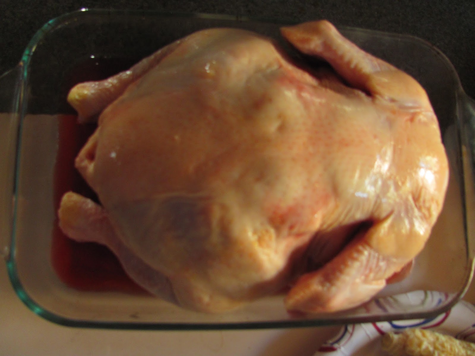
1. Put on gloves. Thoroughly rinse and dry chicken wing. Place it on a plate or tray.


2. Examine the outside skin tissue. Record at least 4 observations of the chicken skin. (1 point)

  • The skin appears to be loose.
  • There are visible hair/feather follicles that make the surface of the skin lumpy.
  • In addition to hair/ follicle bumps, there are other uneven clumps under the epidermal layer of the skin. 
  • It is white in color.

3. Using scissors, knife, and tweezers, work slowly and carefully to cut the skin and peel it away from the muscle below. Notice the clear connective tissue that holds the skin to the muscles. The probe may be the most effective tool. As you peel off the skin, you may need to cut away some connective tissue.

4. Record at least 4 observations of connective tissue. (1 point)

  • The connective tissue is loosely attached to the skin.
  • It also connected to the muscle.
  • It appears to be very fragile.
  • It is transparent. 
5. Take a photo (.5 point)


6. What specific type of connective tissue is this? (.5 point)

  • It has all of the classifications of loose connective tissue. 

7. Observe the yellowish clumps of fat tissue found outside the skin. Record at least 2 observations of the fat. (.5 point)

  • The fat feels tough, as of in a solid form. (Suturated)
  • It's located sporadically across the surface of the skin. 

8. What is the biological term for the type of cells that store fat? (.5 point)

  • The biological name for this type of the fat cells is adipocytes.

9. Name at least 2 functions of this fat. (1 point)

  • It is an energy storage.
  • It also provides an insulation. 

10. Observe bundles of muscle tissue surrounding the bones. Separate the bundles of muscles by separating them out with your fingers. Begin by inserting your thumb into the muscle by pushing through the connective tissue covering the muscle. It will give way at the natural separations between the muscle bundles.


11. Viewing through a magnifying tool, if you have one, (but still do if you don’t) describe 2 characteristics of the arrangement of the muscle bundles as you see them here. (.5 points)

  • Each muscle bundle is enclosed in connective tissue. (Fascia) 
  • Each one is connected to the joints by an individual tendon and to the bone by the ligament.  

12. Using your textbook or another reference, sketch a representation of the muscles starting with the muscle cell and ending with the whole muscle. Cite your source! (3 points)


http://www.student.loretto.org/humanbiology/BioLinks/chp4/slide11.htm


13. What type of muscle tissue are you viewing? (.5 point)

  • This is a skeletal muscle.

14. Name the function of this type of muscle tissue. (1 point)

  • Its function is to allow the movement of the bones. 

15. Name 2 two characteristics of this muscle tissue. (.5 point)

  • This muscle can contract, upon receiving the signal.
  • It also can relax.

16. Nerves are thin, threadlike, white strands found between the muscle and the nearest bone. Remove a single muscle by cutting the tendons and peeling the muscle away from the bone. Look for the nerve in your specimen. The texture is much different from a tendon or bone. It is rather slippery. Did you find one? (.5 points)
  • I did!
17. Take a photo of the muscle and nerve or only the muscle, if you did not find the nerve. (.5 point)


18. The strong, shiny, white cords of tendons hold the muscle to the bones. Some of these tendons pulled away from the bone as you separated the muscle bundles.

Observe with a magnifying tool, if you have one, describe, and sketch

a. the attachment of a tendon to muscle. (.5 point)
  • Thin, tough, white-ish Tendon becomes more and more like the muscle, it branches out with numerous strands, changes the color,  and completely merges with the muscle itself. 
b. the attachment of a tendon to the bone. (.5 point)
  • Tendon extends its thin fibers and attaches itself to the bone. The connection is strong and precise. 


19. Take a photo. (.5)


20. Cut across the tendons at the elbow and peel back toward the carpal joint as if you were peeling a banana. Observe the numerous tendons and pull the freed muscles down and away from the bone. Don't cut any ligaments that attach bone to bone.

21. Take a photo. (.5)


"Dissection of the Chicken Wing & Comparison to Human Arm." We Love Teaching. n.d. Web. 7 Feb. 2015.


22. Look closely at the ligaments with a magnifying tool. Describe 2 differences in appearance between tendons and ligaments. (.5 point)

  • Tendon appears to be "stringy" and it is attached to the muscle.
  • Ligaments look more like an adhesive tape, they hold bones together. 

23. What type of connective tissue composes the ligaments? (.5 point)

  • Ligaments are composed of tough connective tissue.

24. Remove all remaining muscle to expose the bones of the chicken wing.

25. Take a photo. (.5)


26. Bend the elbow. Refer to pages 113-115 in your textbook and answer these questions.

a. What type of joint is this? (.5 point)

  • This is a synovial type of joint.

b. What type of movement is being demonstrated? (.5 point)

  • This joint represents the "hinge" type and it demonstrates the movement in one plane.

27. Cut into the elbow joint and separate the ulna and radius from the metacarpals. Observe the shiny white layer covering the ends of the bones. Name this covering according to its primary tissue and specific type. (1 point)

  • This white layer is hyaline cartilage, it is a transition tissue, and it is a specialized type. 

28. Describe the texture of the ends of the bones at the joint. (.5 point)

  • The texture feels tough but moist at the same time, it is very smooth. 

29. Name three functions of bone. (.5 point)

  • Bones provide protection and the shape of our structure, and they provide us with an ability to move. 
  • They contain the bone marrow, where the stem cells produce red and white blood cells. 
  • They also provide storage for the minerals and fats. 

30. If you could see inside the bone. What soft material would you find? Do not break the raw chicken bone. There is danger from bone fragments flying out. (.5 point)

  • Inside the bone is a soft material called the bone marrow. 

31. Name three specific types of cells present here. (1.5 points)
  • Bone marrow contains two types of stem cells: hemopoietic (which can produce blood cells) and stromal (which can produce fat, cartilage, and bone).
  • There are two types of bone marrow: red marrow (also known as myeloid tissue) and yellow marrow.
  • Red blood cells, platelets, and most of white blood cells arise in the red marrow; some white blood cells develop in the yellow marrow.
  • The color of yellow marrow is due to the much higher number of fat cells.

  • Osteoblast, osteoclast, and osteocyte cells also inhibiting inside of the bone cavity. 

32. Explore and examine other parts.

33. Dispose of materials. Using warm water and soap thoroughly wash all tools and materials, including your hands and the surface you worked upon.


Now that I am done with this lab, everything is cleaned up and the chicken is also done, it is time to eat! 
Bon appetit!


Sources:


"Structure of a Muscle (1 of 4)." Structure of a Muscle (1 of 4). Web. 1 Oct. 2015. <http://www.student.loretto.org/humanbiology/BioLinks/chp4/slide11.htm>.


ScienceDaily. ScienceDaily. Web. 1 Oct. 2015. <http://www.sciencedaily.com/terms/bone_marrow.htm>. Note: The above text is excerpted from the Wikipedia article "Bone marrow", which has been released under the GNU Free Documentation License.

Friday, September 18, 2015

Week 4. Tissues, Skin, Membranes Lab.

Part 1
Tissues Identification and Characterisation.


EPITHELIAL  TISSUE


This is epithelial tissue because the cells are organized and have a structure. They form the layers and have distinctive shapes: squamous, cuboidal, and columnar.


CONNECTIVE TISSUE


This is connective tissue because it have  a few living cells. It does have fibers, fat cells, and ground substance.

MUSCLE TISSUE


This is muscle tissue because it has long strands of muscle fibers which are long and thin. Also, the stands are usually positioned parallel to each other.

NERVOUS TISSUE


This is nervous tissue because it has a large (in comparison to other cells) body, it has the dendrites (extensions that spread out from the cell and receive the signals) and axon, (that transmits the signals).


Part 2.
Tissue Membrane

MUCUS


Part 3.
Body Position and Directional Panes.
a)
https://www.google.com/search
b)   My friend have a blister on the bottom (inferior) of her foot. In addition, it is located on the rear end of her foot (posterior). So, I would say, that she have a blister on her heel.


Part 4.
Skin Structure and Function.


1.  Duration of the sensation: about 4 seconds. 
2.  At a different location, the duration of sensation is the same, about 4 seconds. 
3/4.  After placing three more coins on the same location, the pressure sensation does return, but duration of it lasts for about 4-5 seconds. 
( i had an extra subject present for this experiment, but she didn't last long, so impatient   :) )


5.  I have fouond this simple video on how our skin receives its sensetion. 

Our sense of touch is controlled by a huge network of nerve endings and touch receptors in the skin known as the somatosensory system. This system is responsible for all the sensations we feel - cold, hot, smooth, rough, pressure, tickle, itch, pain, vibrations, and more. Within the somatosensory system, there are four main types of receptors: mechanoreceptors, thermoreceptors, pain receptors, and proprioceptors.

A touch receptor is considered rapidly adapting if it responds to a change in stimulus very quickly. Basically this means that it can sense right away when the skin is touching an object and when it stops touching that object. However, rapidly adapting receptors can't sense the continuation and duration of a stimulus touching the skin (how long the skin is touching an object).
This is why after about 4 or 5 seconds, we can't feel the couins on our skin. 


Appendages of the skin.


Work cited:

"Nose - Google Search." Nose - Google Search. Web. 18 Sept. 2015. <https://www.google.com/search?q=nose&rlz=1C1GGGE_enUS594US614&es_sm=122&source=lnms&tbm=isch&sa=X&ved=0CAcQ_AUoAWoVChMIp4uE17WByAIVwZQNCh3AuA8r&biw=1435&bih=744#tbm=isch&q=mucous glands in nose&imgrc=melaihEOHy_MQM:>.


 "Skin Cross Section - Google Search." Skin Cross Section - Google Search. Web. 18 Sept. 2015. <https://www.google.com/search?q=skin cross section&rlz=1C1GGGE_enUS594US614&espv=2&biw=1435&bih=744&tbm=isch&tbo=u&source=univ&sa=X&ved=0CBwQsARqFQoTCMGpirm8gcgCFQqPDQod_c8D9A#imgrc=OMUa_yxlDpIBzM:>. 


 "Sense of Touch." Skin & Sense of Touch. Home Science Tools. Web. 18 Sept. 2015. <http://www.hometrainingtools.com/a/skin-touch>. 

Friday, September 11, 2015

Week 3. The Microscope, Cells, and Organelles Lab.

Part 1 
 The microscope magnification.


                             Lens mag.                                Ocular mag.                    Total mag.
a. Scanning                4x                    multiplied by        10x                =                 40x

b. Low Power          10x                    multiplied by        10x               =                100x

c. High Power          40x                   multiplied by         10x                =               400x

d. Oil immersion    100x                   multiplied by         10x                =             1000x


Part 2
Virtual Microscope
                         
    ONION ROOT  

                        
4x
10x

40x

100x
Onion root, under magnification of 100x, has the visible cells with their nucleus (dark blue), cytoplasm (lighter blue substance surrounding the nucleus), and cell walls (which are better viewed at mag. 40x).

CHEEK CELLS SMEAR

4x

10x
40x
100x


In the cheek cell image, with mag. of 100x., I can see the nucleus, cytoplasm, and organelles (in the 40x mag. image.)

Answers to part 2 questions.


1. What structure separates the contents of the nucleus from the cytoplasm?

The double-layered membrane, called the nuclear membrane, separates the nucleus from the cytoplasm. 

2. What is the function of the nucleoli? Why is this important


The nucleoli are located in the body of the nucleus. As the nucleus is the "brain" of the cell, the nucleolus (numerous nucleoli) could loosely be thought of as the brain of the nucleus. The nucleolus takes up around 25% of the volume of the nucleus.This structure is made up of proteins and ribonucleic acids (RNA). Its main function is to rewrite ribosomal RNA (rRNA) and combine it with proteins. This results in the formation of incomplete ribosomes. It produces the energy.


3. Other than the nucleoplasm and the nucleoli, what are three other materials found in a nucleus?

The three materials that found in a nucleus, besides nucleoplasm and nucleoli, are DNA, RNA, and ribosomal proteins. 

Operating the Microscope.

4. Which objective should be in place when you start?

The smallest magnification objective should be in place when we start, usually it is the scanning one, with mag. of 4x.


5. Which focusing knob do you first use when you begin looking at a slide?

When we begin looking at a slide, we first use the coarse knob to focus on it. 

6. What do you adjust if you can see two overlapping circles with part of the object in each circle?

To eliminate the two overlapping circles when looking at the slide, we have to adjust the oculars by moving them from side to side, until the desired one circle, with proper focus. 

7. How do you move the slide on the stage?

To move the slide on the stage, we have to adjust the two "stage manipulator knobs" to reposition the slide one moves it left-right and the other  moves it up-down, closer to the center with the light. 

8. What two devices can be maneuvered to increase or decrease light intensity?

To increase or decrease the light intensity, we can maneuver the rheostat and/or iris diaphragm lever. 

9. What is the name of the highest magnifying objective?

The highest magnifying objective is called "oil immersion".

10. Which objective allows you to see the largest area of the object that you are viewing?

To view the largest area of the object, we have to use the scanning objective, the one with the magnification of 4x.  


Part 3

Functions of cells and organelles.

Procedure
  1.  Pour 8 ounces tap water into each of two cups or glasses. Add 3 teaspoons salt to one of the cups and label it "Salt Water" with a piece of masking tape. Label the other cup "Fresh Water".
  2.    Measure the length and greatest width of both pieces/carrots and record this measurement in Table 1 below.


  3.    Tightly tie a piece of string around each carrot.
  4.   Set one carrot/carrot piece down into the “Fresh Water” cup and the other down into the “Salt Water” cup.
  5.  Take a photo of the two containers with their contents.

  6.  Allow the carrots to sit in the cups for 24 hours.
  7.  Ask yourself what will happen to both carrots in this experiment in terms of changes in length and diameter and why you think this will happen.  Record your predictions and rationale.

  • I think that the carrot in the salt water solution will shrink slightly compared to the carrot in the fresh water. 
  • Freshwater carrot will remain the same size because of the balanced concentration of the inside and the outside fluid. 

  1. After 24 hours, remove the carrots and examine the tightness of the strings. Re-measure the carrots and record the findings in Table 1 below.

 Remove carrots from cups and take a photo next to each other outside of the containers.
 Record more observations.


a.       Note if there are any differences in the tightness of the strings and the textures for both carrots. Observe which type of water caused an increase in cell size.
b.      Observe which type of water caused a decrease in cell size.
c.       How did the actual results in the measurements compared to your predictions?

After taking it out of the water, the salt water carrot is visibly smaller and the string tied around it was loose. It also looks somewhat "wrinkled", or a better word would be shriveled. 
The salt water caused the carrot to decrease in its size. .
It looks like my prediction was true to the actual results. The carrot in the salt water did shrink and became smaller in size. 

Remember science does not prove anything to be true. Through experimentation, results can illustrate processes and these processes may be used to predict similar events in similar or different circumstances.


What is osmosis? Include these elements in your explanation:

Osmosis is the spontaneous net movement of solvent molecules through a semi-permeable membrane into a region of higher solute concentration, in the direction that tends to equalize the solute concentrations on the two sides. It may also be used to describe a physical process in which any solvent moves across a semipermeable membrane (permeable to the solvent, but not the solute) separating two solutions of different concentrations.

What is moving? Why?

When two different solutions have different molecular density concentrations, the diffusion occurs. 


The molecules with a higher concentration will be moving towards the area with the less concentration, to "balance" the solution's consistency. 


 What does not move? Why?

The molecules will not be moving if the concentration is neutral. Like adding water to water will "mix" the water molecules, but will not unbalance the total consistency.

 How is this important for the health of cells?

When cells encounter the undesirable substance (like a sodium), they will not only work very hard to maintain their inner balance but also will lose their intracellular fluid due to "overworking", which will result in the "shrinking" of the cell and in extreme cases, the complete destruction of it. (the cell will die)


Table 2. Starting and Ending Carrot Measurements


Length at Start of 24 Hours
Length at End of 24 Hours
Width at Start of 24 Hours
Width at End of 24 Hours
Freshwater Carrot
 2 inches

2 inches
 .75 inch
.75 inch

Saltwater Carrot
 2 inches

1.75 inch
 .75 inch
.5 inch


Sources:


 "Function of Nucleolus." Function of Nucleolus. Web. 11 Sept. 2015.   <http://www.softschools.com/science/biology/function_of_nucleolus/>. 


Wikipedia. Wikimedia Foundation. Web. 14 Sept. 2015. 


Wednesday, September 2, 2015

Week 2 Acid-Base and Molecules Lab

Part 1
Acids and Bases
Types of protein

a. ACTIN (skeletal and muscular component)

b. TUBULIN (cellular structure)

c. HEMOGLOBIN (transports oxygen)

d. KERATIN (structural support)

e. IMMUNOGLOBIN (immune system support)

f. ALBUMIN (development of the embryo)

g. MYOSIN ( muscular contraction)

Reaction with the acid. 

Let's look at the albumin.

Because it is a protein that responsible for a development of an embryo, my example for the purpose of visualization, will be an egg. Adding an acid to the protein, will break hydrogen bond between protein's molecules. With that breakage, the structure of the uniform protein becomes so disorganized, it will experience the process of denaturing. When we beat the liquid egg whites, we actually rearranging the structure of the protein and the result is a foamy,  new bond of protein. The disorganization of molecules also happens when we add an acid, Like a lemon. The restructured protein will lose its potency, and will become a separate structure from the original bond. The egg will begin to "cook", the color will change to white and the consistency will become harder. Heat also will restructure the original bond and will change the consistency of the protein.  Hard boiled egg anyone? 


Part 2

Molecule A
http://riversideonline.com/employees/myhealthylifestyle/newsletter/fructose-sucrose-glucose.cfm?&RenderForPrint=1 
1) Belongs to Sucrose group
2) Common term: table sugar

Molecule B

http://www.indiana.edu/~oso/Fat/SolidNLiquid.html
1) Belongs to Triglycerides group
2)Common term: Saturated fat, or animal fat, or commonly used and loved bacon fat.


Molecule C

http://thenaturalhistorian.com/2013/12/05/ancient-dna-creationism-genome-sequencing/
1) Belongs to Nucleic-Acid group
2) Common term: or as known as - DNA

Molecule D

http://www.precisionnutrition.com/all-about-testosterone
1) Belongs to Testosterone group
2) Common term: Male sex hormone.

Molecule E


http://chemwiki.ucdavis.edu/Textbook_Maps/General_
Chemistry_Textbook_Maps/Map%3A_Lower's_Chem1/07%3A_Solids_and_Liquids/7.3%3A_Hydrogen-Bonding_and_Water
1) Belongs to Proteins
2) Common term: beans, eggs, nuts


Sources

"Actin." Definition of in Biology. Web. 3 Sept. 2015.

"Types and Functions of Proteins - Boundless Open Textbook." Boundless. Web. 3 Sept. 2015.        <https://www.boundless.com/biology/textbooks/boundless-biology-textbook/biological-    macromolecules-3/proteins-56/types-and-functions-of-proteins-302-11435/>.
 
"Sweet Things." Sweet Things. Web. 3 Sept. 2015. <http://riversideonline.com/employees/myhealthylifestyle/newsletter/fructose-sucrose-glucose.cfm?&RenderForPrint=1>. 
        http://riversideonline.com/employees/myhealthylifestyle/newsletter/fructose-sucrose-glucose.cfm?&RenderForPrint=1

"Sweet Things." Sweet Things. Web. 3 Sept. 2015. <http://riversideonline.com/employees/myhealthylifestyle/newsletter/fructose-sucrose-glucose.cfm?&RenderForPrint=1>.          
       http://www.indiana.edu/~oso/Fat/SolidNLiquid.html

"Sweet Things." Sweet Things. Web. 3 Sept. 2015. <http://riversideonline.com/employees/myhealthylifestyle/newsletter/fructose-sucrose-glucose.cfm?&RenderForPrint=1>.

"All about Testosterone: What You Need to Know about the King of Male Hormones. | Precision Nutrition." Precision Nutrition. 22 Feb. 2010. Web. 3 Sept. 2015. <http://www.precisionnutrition.com/all-about-testosterone>.

"7.3: Hydrogen-Bonding and Water." - Chemwiki. 2 Oct. 2013. Web. 3 Sept. 2015. <http://chemwiki.ucdavis.edu/Textbook_Maps/General_Chemistry_Textbook_Maps/Map:_Lower's_Chem1/07:_Solids_and_Liquids/7.3:_Hydrogen-Bonding_and_Water>.

 "7.3: Hydrogen-Bonding and Water." - Chemwiki. 2 Oct. 2013. Web. 3 Sept. 2015. <http://chemwiki.ucdavis.edu/Textbook_Maps/General_Chemistry_Textbook_Maps/Map:_Lower's_Chem1/07:_Solids_and_Liquids/7.3:_Hydrogen-Bonding_and_Water>.